


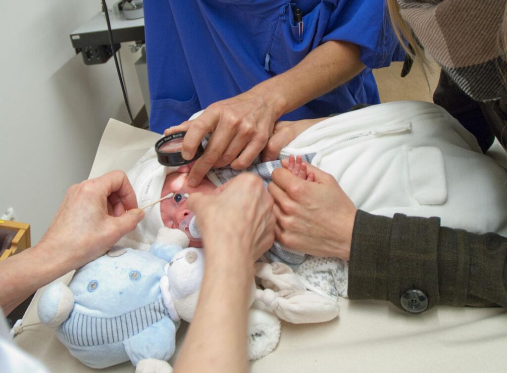
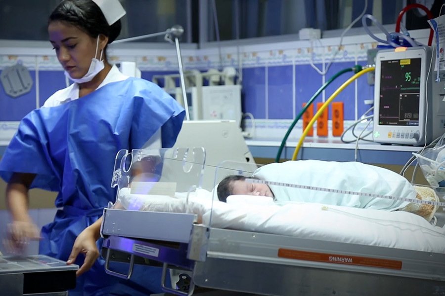
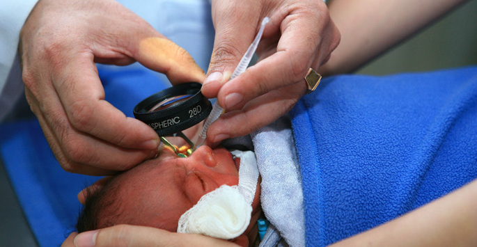
Retinopathy of prematurity (ROP) is a disease that occurs in premature babies. It causes abnormal blood vessels to grow in the retina, the layer of nerve tissue in the eye that enables us to see. This growth can cause the retina to detach from the back of the eye, leading to blindness.
Some cases of ROP are mild and correct themselves, but others require surgery to prevent vision loss or blindness. Surgery involves using a laser or other means to stop the growth of the abnormal blood vessels, making sure they don’t pull on the retina. The inside of the eye, the retina is not fully developed in premature babies. Abnormal blood vessels can develop in such a retina. These abnormal blood vessels can cause internal bleeding and even retinal detachment. This is called Retinopathy of Prematurity (ROP). This condition results in low vision or blindness – both of which are irreversible.
Blood vessels grow from the center of a developing baby’s retina 16 weeks into the mother’s pregnancy, and then branch outward and reach the edges of the retina 8 months into the pregnancy. In babies born prematurely, normal retinal vessel growth may be disrupted and abnormal vessels can develop, which can cause leaking and bleeding in the eye. ROP can stop or reverse itself at any point, so it often resolves as the baby grows. Sometimes, though, the disease may progress to cause scarring, which pulls the retina away from the rest of the eye. ROP has no signs or symptoms. The only way to detect it is through an eye examination by an ophthalmologist.
If admission to the hospital isn’t necessary, you’ll be able to take your child home about an hour after the procedure. Follow-up care for ROP surgery includes giving your child eye drops (to prevent infection) for at least a week.
To make sure the eyes are healing properly and that ROP hasn’t returned, eye exams should be scheduled based on instructions from the ophthalmologist. This is usually every 1-2 weeks. For scleral buckling, the ophthalmologist must examine the buckle every 6 months to account for your child’s growing eye. The goal of surgery for retinopathy of prematurity is to stop the progression of the disease and prevent blindness. Although ROP surgery has a good success rate, not all babies respond to treatment. Up to 25% of babies who have ROP surgery might still lose some or all vision.
Keep in mind that for all types of ROP surgery, a degree of your child’s peripheral (side) vision will be lost. And even if the ROP has stopped progressing, vision still can be affected. Since some vision loss and complications can occur, any child who has had ROP surgery should have regular, yearly eye exams well into adulthood.
ROP surgery is used to stop the growth of abnormal blood vessels by focusing treatment on the peripheral retina (the sides of the retina) to preserve the central retina (the most important part of the retina). ROP surgery involves scarring areas on the peripheral retina to stop the abnormal growth and eliminate pulling on the retina.
Since surgery focuses treatment on the peripheral retina, these areas will be scarred and some amount of peripheral vision may be lost. However, by preserving the central retina, the eye will still be able to perform vital functions like seeing straight ahead, distinguishing colors, reading, etc. The most frequently used methods of ROP surgery are:
RETINA SPECIALITY HOSPITAL prides itself in providing the highest standard-of-care in the most cost-effective way to its patients. The staff & doctor of this hospital, believe in personal & patient - friendly services...Read More
Copyright © 2021 Retina Speciality Hospital
Design & Developed By Namastetu
Developed by Namastetu Technologies